Hormonal Pathways in Water Retention

Understanding ADH, aldosterone, and ANP signaling cascades that orchestrate fluid balance.

Hormonal signaling pathways for water retention

Antidiuretic Hormone (ADH)

ADH (vasopressin) is a nine-amino-acid peptide synthesized by the hypothalamus and released from the posterior pituitary gland. ADH represents the primary mechanism for short-term water balance adjustment, responding to changes in plasma osmolarity and blood volume within minutes.

Osmotic Regulation

Osmoreceptors in the hypothalamus continuously monitor plasma osmolarity. Increased osmolarity stimulates ADH release, promoting water reabsorption in renal collecting ducts. This increases plasma volume and dilutes blood osmolarity back toward normal. Decreased osmolarity suppresses ADH release, reducing collecting duct water permeability and allowing water excretion as dilute urine. The osmotic threshold for ADH secretion is approximately 285 mOsm/kg; minimal ADH secretion occurs below this threshold, and maximal secretion occurs at osmolarity above 300 mOsm/kg.

Volume Regulation

Baroreceptors in carotid sinuses and aortic arch also regulate ADH. Significant blood volume decreases stimulate ADH release despite decreased plasma osmolarity, overriding osmotic inhibition. This volume regulation protects against excessive blood loss and hemodynamic collapse. Conversely, blood volume expansion suppresses ADH regardless of plasma osmolarity. The volume threshold requires approximately 10% blood volume change before override of osmotic regulation occurs.
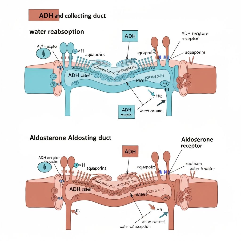
Mechanism of Action

ADH binds to V2 receptors on collecting duct principal cells. Receptor activation increases cAMP, activating protein kinase A, which phosphorylates regulatory proteins. Phosphorylated proteins insert aquaporin-2 water channels into the apical membrane, increasing water permeability. When ADH levels decline, endocytosis internalizes aquaporin-2 channels, reducing water permeability. This reversible channel regulation permits rapid adjustment of water reabsorption in response to body needs.

Aldosterone and Mineralocorticoid Regulation

Aldosterone is a steroid hormone produced by the adrenal cortex zona glomerulosa. Unlike ADH which operates through short-term mechanisms, aldosterone regulates sodium balance through longer-term gene expression changes, requiring hours to manifest full effects.

Stimulation of Aldosterone Secretion

Renin-angiotensin system activation represents the primary stimulus for aldosterone secretion. When renal perfusion pressure decreases, juxtaglomerular cells release renin. Renin cleaves angiotensinogen to angiotensin I; angiotensin-converting enzyme (ACE) produces angiotensin II. Angiotensin II powerfully stimulates aldosterone synthesis and release from zona glomerulosa cells.

Serum potassium directly stimulates zona glomerulosa cells; hyperkalemia triggers aldosterone release independent of renin. This direct mechanism ensures potassium homeostasis, as aldosterone increases urinary potassium excretion.

ACTH provides minor stimulation of aldosterone during stress responses, though this effect is transient despite sustained ACTH elevation.

Aldosterone Mechanism of Action

Aldosterone crosses collecting duct cell membranes and binds to mineralocorticoid receptors in the cytoplasm. The ligand-receptor complex translocates to the nucleus, altering gene transcription. Aldosterone increases expression of epithelial sodium channels (ENaC) and Na-K-ATPase. Enhanced sodium reabsorption creates a negative intraluminal charge, facilitating potassium and hydrogen ion secretion. Water follows reabsorbed sodium osmotically, expanding extracellular fluid volume.

Atrial Natriuretic Peptide (ANP)

ANP antagonizes sodium and water retention, promoting urinary sodium and water excretion. ANP is synthesized and released by cardiac atrial myocytes in response to atrial stretch triggered by increased blood volume.

ANP Actions

ANP binds to natriuretic peptide receptors (NPR-A) on collecting duct cells and vascular smooth muscle. In the kidney, ANP increases glomerular filtration rate by dilating afferent arterioles and constricting efferent arterioles, increasing glomerular filtration pressure. Additionally, ANP inhibits sodium reabsorption in the collecting duct and inhibits aldosterone synthesis. In blood vessels, ANP causes vasodilation, reducing blood pressure. ANP also inhibits renin and ADH secretion, providing multiple coordinated effects reducing blood volume and normalizing blood pressure.

The Renin-Angiotensin-Aldosterone System (RAAS)

The RAAS represents a critical mechanism coordinating blood pressure, blood volume, and electrolyte balance. Multiple factors activate this system, triggering a coordinated response restoring renal perfusion pressure.

RAAS Activation and Actions

Decreasing renal perfusion pressure stimulates juxtaglomerular cell renin release. Decreased serum sodium, increased potassium, and sympathetic nervous system activation also promote renin secretion. Renin initiates the cascade producing angiotensin II, which causes systemic vasoconstriction increasing blood pressure and preferentially dilates efferent arterioles, increasing glomerular filtration pressure. Angiotensin II directly stimulates aldosterone synthesis, promoting sodium and water reabsorption. The combined effects restore blood volume and pressure.

Negative Feedback Control

Restored blood pressure inhibits further renin secretion through baroreceptor signaling. Increased sodium delivery to the macula densa suppresses renin secretion. Elevated angiotensin II inhibits renin secretion through direct negative feedback. ANP suppresses renin secretion, aldosterone synthesis, and ADH release. These multiple feedback mechanisms prevent excessive RAAS activation and normalize blood volume and pressure.

Coordination of Hormonal Systems

Water and electrolyte balance requires coordinated interaction between multiple hormonal systems. These systems respond to overlapping stimuli and often provide redundant control, ensuring homeostatic stability.

Integrated Responses

Hypovolemia (low blood volume) triggers simultaneous ADH and aldosterone activation. ADH promotes water reabsorption, restoring osmolarity while increasing blood volume. Aldosterone promotes sodium and water reabsorption through ENaC activation. RAAS activation from decreased renal perfusion pressure amplifies these effects. ANP secretion decreases, removing its inhibitory influence. The combined effect rapidly restores blood volume and pressure.

Hypervolemia (excess blood volume) suppresses ADH and aldosterone while stimulating ANP. Decreased ADH reduces collecting duct water permeability, permitting water excretion. Decreased aldosterone reduces sodium reabsorption. Increased ANP promotes sodium and water excretion while inhibiting aldosterone and ADH. Renal perfusion pressure increases, suppressing renin secretion. The coordinated response eliminates excess fluid.

Hypernatremia (elevated serum sodium) stimulates ADH secretion despite potential volume expansion. The osmotic stimulus overrides volume signals, promoting water reabsorption to dilute blood sodium. Simultaneously, decreased aldosterone secretion (from decreased renin) reduces sodium reabsorption, helping normalize sodium concentration.

Hyponatremia (decreased serum sodium) suppresses ADH, permitting water excretion. Simultaneously, increased aldosterone from RAAS activation promotes sodium reabsorption. ANP secretion increases, further promoting sodium excretion. The coordinated response restores sodium concentration.

Clinical Relevance of Hormonal Dysregulation

Dysfunction in any component of these hormonal systems can produce pathological fluid retention or excessive fluid loss. Understanding these mechanisms provides context for recognizing how various conditions affect everyday fluid balance experiences.

Syndrome of inappropriate ADH (SIADH) involves excessive ADH secretion, promoting inappropriate water reabsorption and hyponatremia. Central diabetes insipidus results from insufficient ADH production, causing polyuria and hypernatremia. Nephrogenic diabetes insipidus reflects collecting duct insensitivity to ADH, similarly producing polyuria.

Primary aldosteronism causes excessive sodium and water reabsorption, leading to hypertension and hypokalemia. Addison's disease (adrenal insufficiency) produces aldosterone deficiency, causing sodium and water loss. Conn syndrome specifically involves aldosterone-producing adenomas, causing selective hyperaldosteronism.

Heart failure stimulates RAAS activation and ADH release despite volume expansion, reflecting baroreceptor dysfunction and promoted sodium-water retention contributing to peripheral edema and pulmonary congestion.

Return to Blog